
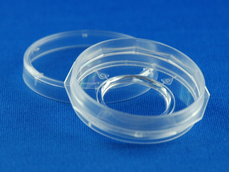
Imaging Dish
35 mm petri dish with central
18 mm diameter cover glass bottom
Order information
Article number | Cover glass thickness | Unit/Bag | Dishes/Sales Unit | Lid | Sterile |
5160-30 | 1.0, 145 µm | 2 / Bag | 30 / Box | + | + |
5160-150 | 1.0, 145 µm | 2 / Bag | 150 / Box | + | + |
6160-30 | 1.5, 170 µm | 2 / Bag | 30 / Box | + | + |
6160-150 | 1.5, 170 µm | 2 / Bag | 150 / Box | + | + |
6160-30 *New* | 1.5H, 170 µm high precision glass | 2 / Bag | 30 / Box | + | + |
6160-150 *New* | 1.5H, 170 µm high precision glass | 2 / Bag | 150 / Box | + | + |
-
Low skirt format: ideal for immersion microscopy
-
Robust format: stability against focal shift during temperature changes and time lapse studies
-
12 notch grip zone: easy grip for dish and lid
-
Orientation marks: align dish on microscope stage
-
Imaging reservoir: reduced volume for staining and labeling
-
Tissue Culture Surface treatment: improved cell spreading, adhesion and growth
- *New*: Imaging Dish with a high precision glass for high-resolution analyses, e.g. for STED or STORM super resolution
Life Cell Imaging, Fluorescence-Correlation Spectroscopy (FCS), Confocal Laser Scanning Microscopy (LSM), Total Internal Reflection Fluorescence (TIRF), Differential Interference Contrast (DIC/Nomarski), Fluorescence Resonance Energy Transfer (FRET), Fluorescence Recovery after Photo bleaching (FRAP), Weakly fluorescent stains / proteins, Fluorescence In-Situ Hybridization (FISH), Immune histology
The Imaging Dish products are our entry solution for high resolution microscopy of living and fixed cells. The bottom of the 35 mm petri dish is made from cover glass. The central cover glass area has a diameter of 18 mm. The level of the cover glass is 2 mm lower than the basement level of the surrounding polystyrene dish. Due to this feature it is possible to concentrate the cells during the seeding on the glass surface.
Several features support a user friendly manipulation of the dish. The grip zone of the dish body is increased and allows a safe picking of the dish. The lid contains spacers for good ventilation. For cell manipulations the lid can easily be removed. The dishes are delivered steril in bags of two dishes.
The focus level of the glass area is placed only 400 µm above the basement line of the dish body. High quality cover glasses made from borosilicate glass are used. Variants with cover glass thickness 1.0 and 1.5 (145 µm and 170 µm ± 15µm respectively) are offered. These glass bottoms provide an excellent planarity and focus control and superior imaging qualities.
The article Imaging Dish is now also available with a “high-precision glass”. These are used for high-resolution analyses, e.g. for STED or STORM super resolution.
The macroscopic orientation of the dishes is supported by four marks (N-O-S-W). The twelve corner grip zone of the dishes enables an easy orientation and positioning of the dies if combined with our Imaging Dish Stage Frame. Therefore the same orientation of the dishes can be established even if the dish has to be removed from the stage in between two observation sessions.
|
Cover glass properties |
|
|
Thickness |
1.0, 145 µm +/- 10% 1.5, 170 µm +/- 10% 1.5H, 170 µm +/- 3% |
|
Glass type |
borosilicate, hydrolytic class I |
|
Refractive Index |
1,52 |
|
Abbe’s number |
50 |
|
Light transmission @ 310 nm |
> 85% |
|
Light transmission @ 360 nm |
> 90% |
|
Surface quality |
TC (Tissue Culture Quality) |
TC= Tissue Culture, amine coated surface, suitable for most adherend cell lines (e.g. HEK U293, HepG2, MCF-7). Sensitive cell lines (e.g. PC-12) and some primary cell types benefit from additional surface coatings with ECM proteins or equivalent coatings.
|
Article number |
5160/6160/6170 |
|
Outer dish diameter |
35 mm |
|
Diameter cover glass area |
18 mm |
|
Dish height |
9,4 mm |
|
Distance dish bottom rim to focus plane |
400 µm |
|
Planarity |
< 5 µm |
|
Total dish volume |
7 ml |
|
Suggested working volume |
2 ml |
|
Suggested cell seeding volume |
750 μl |