
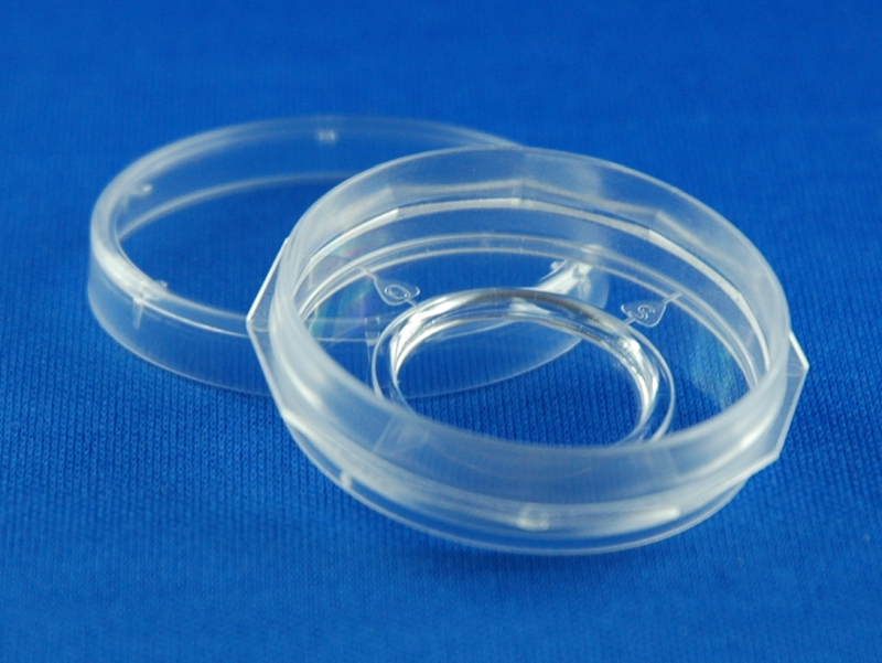
Imaging Dish µGrid
35 mm petri dish with central
18 mm diameter cover glass bottom
with engraved µGrid
Order information
Article number | Cover glass thickness | Unit/bag | Dishes/ Sales unit | Lid | Sterile |
7160-30 | 1.5, 170 µm | 2 / bag | 30 / box | + | + |
7160-168 | 1.5, 170 µm | 2 / bag | 168 / box | + | + |
Laser engraved µGrid for cell localization
Low skirt format: ideal for immersion microscopy
Robust format: stability against focal shift during temperature changes and time lapse studies
12 notch grip zone: easy grip for dish and lid
Orientation marks: align dish on microscope stage
Imaging reservoir: reduced volume for staining and labelling
Tissue Culture Surface treatment: improved cell spreading, adhesion and growth
Cytometry, Cell localization and re-localization, Life Cell Imaging, Fluorescence-Correlation Spectroscopy (FCS), Confocal Laser Scanning Microscopy (LSM), Total Internal Reflection Fluorescence (TIRF), Differential Interference Contrast (DIC/Nomarski), Fluorescence Resonance Energy Transfer (FRET), Fluorescence Recovery after Photo bleaching (FRAP), Weakly fluorescent stains / proteins, Fluorescence In-Situ Hybridization (FISH), Immune histology
Imaging Dish µGrid are equivalent to the Imaging Dish 1.5 (170 µm cover glass bottom) but the cover glass carries in addition a micro grid. This µGrid located in the focus plane of the cover glass. The micro grid supports localization and re-localization of the Region Of Interest (ROI) even if no computer assisted motor stage is available. The grid is oriented towards the macroscopic orientation marks of the Imaging Dish. When the Imaging Dish µGrid is combined with the Imaging Dish Stage Frame it is easily possible to navigate to the ROIs at different time points, even if the Imaging Dish is removed from the microscope stage in between.
The µGrid is made by precise laser marking. The single grid lines have a width of 3 µm, a length of 9 mm and a depth of 0,3 µm. In wide field and phase contrast illumination the lines are visible but do not influence cell attachment or orientation. In fluorescence mode the grid lines are not visible and do not cause artefacts during the image acquisition.
The focus level of the glass area is placed only 400µm above the basement line of the dish body. High quality cover glasses made from borosilicate glass are used. These glass bottoms provide an excellent planarity and focus control and superior imaging qualities.
The macroscopic orientation of the dishes is supported by four marks (N-O-S-W). The combination of engraved micro grids to support microscopic orientation to retrieve the region of interest (ROI) with the twelve corner grip zone of the dishes enables an easy orientation and positioning of the dishes if combined with our Imaging Dish Stage Frame. Therefore the same orientation of the dishes can be established even if the dish has to be removed from the stage in between two observation sessions.
Thickness | 1.5, 170 µm +/- 10% |
Glass type | borosilicate, hydrolytic class I |
Refractive Index | 1,52 |
Abbe’s number | 50 |
Light transmission @ 310 nm | > 85% |
Light transmission @ 360 nm | > 90% |
Surface quality | TC (Tissue Culture Quality) |
TC= Tissue Culture, amine coated surface, suitable for most adherend cell lines (e.g. HEK U293, HepG2, MCF-7). Sensitive cell lines (e.g. PC-12) and some primary cell types benefit from additional surface coatings with ECM proteins or equivalent coatings.
Article number | 7160 |
Outer dish diameter | 35 mm |
Diameter cover glass area | 18 mm |
Dish height | 9,4 mm |
Distance dish bottom rim to focus plane | 400 µm |
Planarity | < 5 µm |
Total dish volume | 7 ml |
Suggested working volume | 2 ml |
Suggested cell seeding volume | 750 μl |