
Imaging Plate FC
Black multiwell plate with fluorocarbon film bottom
Order information
Article number | Wells/Plate | Plates/bag | Plates/Sales Unit | Lid | Sterile |
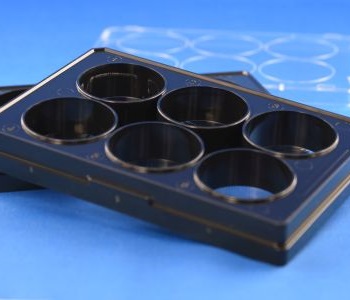
3221-20 | 6 | 1/bag | 20/box | + | + |
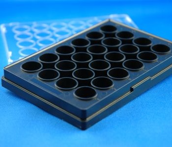
3231-20 | 24 | 1/bag | 20/box | + | + |
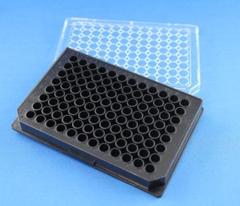
3241-20 | 96 | 1/bag | 20/box | + | + |
Gas permeable bottom
UV light transmission
Low skirt design, complete area access with immersion objectives
Tissue Culture surface
Excellent planarity
Life cell imaging, immunohistochemistry, super high resolution microscopy, laser scanning microscopy, high content analysis, hypoxia studies, phototoxicity studies
The Imaging Plate FC is a black multiwell plate compliant to the SBS (Society for Biomolecular Screening) standard format with a bottom made from a 25 µm fluorocarbon film (FC). During the manufacturing the thin film is stretched around each individual well bottom to provide tension and planarity. Because of the thin bottom and material properties the plates show excellent light transmission rates even for UV-A and UV-B light. The intrinsic fluorescence of the material is much lower if compared to conventional polystyrene bottoms with significant reduction of background signals. Furthermore the film bottom enables a high gas transfer through the film. Oxygen supply and equilibration with the atmosphere are achieved directly through the plate bottom. The plate bodies are made from polystyrene and tightly bonded to the FC film. The Imaging Plate FC can be used for high resolution Live Cell Imaging. In addition direct fixation and staining within the plates is possible (formaldehyde, glutaraldehyde, alcohols and acetone can be used). Acetone should not be applied more than 10 minutes because the plate body is made from polystyrene and subsequent to the material reaction the film bottom may detach. The Imaging Plate FC can be used between -20°C and 50°C. Due to the thin film bottom the application of high resolution immersion objectives with high numerical aperture is possible. For optimal focus control when objectives with 40x magnification and higher the use of objectives with correction rings or collars is recommended. Water, oil and glycerin can be used as immersion media.
Note: All polymer plate bodies react to temperature shifts with dimensional changes. Please take care that the plate and its environment have equal temperature during microscopy to minimze focal shifts and reduce refocusing.
Thickness | 25 µm +/- 10% |
Film type | high performance fluorocarbon film |
Refractive index | 1,34 |
Abbe’s number | 70 |
Light transmission @ 240 nm | > 85% |
Light transmission @ 300 nm | > 90% |
Oxygen transfer capacity | >6300 cm3/(m2*d* bar) |
Carbon dioxide transfer capacity | >7000 cm3/(m2*d* bar) |
Thermal transfer capacity | 0.01 mW/K |
Surface quality | TC (Tissue Culture Quality) |
TC= Tissue Culture, amine coated surface, suitable for most adherend cell lines (e.g. HEK U293, HepG2, MCF-7). Sensitive cell lines (e.g. PC-12) and some primary cell types benefit from additional surface coatings with ECM proteins or equivalent coatings
Article number | 3221 | 3231 | 3241 |
Format | 6 well | 24 well | 96 well |
Plate length | 127,76 mm | 127,76 mm | 127,76 mm |
Plate width | 85,48 mm | 85,48 mm | 85,48 mm |
Plate height | 15 mm | 15 mm | 14,35 mm |
Depth per well | 14,6 mm | 14,6 mm | 13,95 mm |
Well diameter (focal plane) | 32 mm | 13,2 mm | 6 mm |
Distance well center to well center | 36 mm | 18 mm | 9 mm |
Distance center of well A1 to longitudinal plate border | 24,74 mm | 15.74 mm | 11.24 mm |
Distance center of well A1 to short plate border | 27,88 mm | 18,88 mm | 14,38 mm |
Distance lower plate level (skirt rim) to focal plane | 400 µm | 400 µm | 400 µm |
Planicity per well | < 10 µm | < 10 µm | < 10 µm |
Planicity per plate | < 50 µm | < 50 µm | < 50 µm |
Total volume per well | 12112 µl | 1880 μl | 428 µl |
Suggested working volume peer well | 2000-4000 µl | 500-1000 μl | 100-200 µl |