
Imaging Plate CG
Black multiwell plate with cover glass bottom
Order information
Article number | Wells / Plate | Coverglass thickness | Plates/ bag | Plates/ Sales Unit | Lid | Sterile |
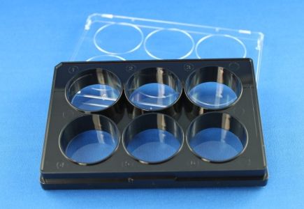
5221-20 | 6 | 145 µm (1.0) | 1 / bag | 20 /box | + | + |
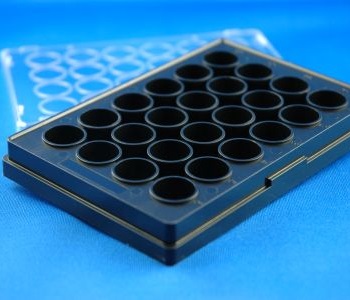
5231-20 | 24 | 145 µm (1.0) | 1 / bag | 20 / box | + | + |
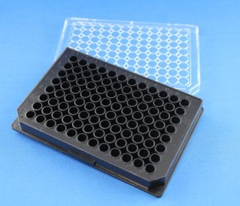
5241-20 | 96 | 145 µm (1.0) | 1 / bag | 20 / box | + | + |
5222-20 | 6 | 170 µm (1.5) | 1 / bag | 20 / box | + | + |
5232-20 | 24 | 170 µm (1.5) | 1 / bag | 20 / box | + | + |
5242-20 | 96 | 170 µm (1.5) | 1 / bag | 20 / box | + | + |
Low skirt design, complete area access with immersion objectives
Tissue culture surface borosilicate cover glass
Excellent planarity
Life cell imaging, immunohistochemistry, super high resolution microscopy, fluorescence correlation spectroscopy, laser scanning microscopy, high content analysis
The Imaging Plate CG is a black multiwell plate compliant to the SBS (Society for Biomolecular Screening) standard-format with a cover glass bottom made from borosilicate glass. The plate bodies are made from polystyrene and tightly bonded to the cover glass. The Imaging Plate CG can be used for high resolution Live Cell Imaging. In addition direct fixation and staining within the plates is possible (formaldehyde, glutaraldehyde, alcohols and acetone can be used). Acetone should not be applied more than 10 minutes because the plate body is made from polystyrene and subsequent to the material reaction the cover glass may detach. Imaging Plate CG can be used between -20°C and 50°C. Due to the cover glass bottom the application of high resolution immersion objectives with high numerical aperture is possible. Water, oil and glycerin can be used as immersion media.
Cell adhesion, spreading and distribution are supported by our approved plasma surface modification.
The cover glass is still the best performing object carrier for high resolution light microscopy. Imaging Plate CG products are equipped with a bottom made from 145µm or 170µm thick borosilicate glass. To support cell adhesion, spreading and equal distribution the glass is plasma modified to increase the amount of amino groups.
All Imaging Plates have a black plate body to suppress cross signals and to prevent excitation of regions out of interest in epifluorescence applications.
All Imaging Plates are single packed and shipped with a lid.
All lids contain condensation rings and spacers for optimal ventilation.
Note: All polymer plate bodies react to temperature shifts with dimensional changes. Please take care that the plate and its environment have equal temperature during microscopy to minimize focal shifts and reduce refocusing.
Thickness | 1.0, 145 µm +/- 10% 1.5, 170 µm +/- 10% |
Glass type | borosilicate, hydrolytic class I |
Refractive Index | 1,52 |
Abbe’s number | 50 |
Light transmission @ 310 nm | > 85% |
Light transmission @ 360 nm | > 90% |
Surface quality | TC (Tissue Culture Quality) |
TC= Tissue Culture, amine coated surface, suitable for most adherend cell lines (e.g. HEK U293, HepG2, MCF-7). Sensitive cell lines (e.g. PC-12) and some primary cell types benefit from additional surface coatings with ECM proteins or equivalent coatings.
Article number | 5221 / 5222 | 5231 / 5232 | 5241 / 5242 |
Format | 6 well | 24 well | 96 well |
Plate length | 127,76 mm | 127,76 mm | 127,76 mm |
Plate width | 85,48 mm | 85,48 mm | 85,48 mm |
Plate height | 15 mm | 15 mm | 14,35 mm |
Depth per well | 14,6 mm | 14,6 mm | 13,95 mm |
Well diameter (focal plane) | 32 mm | 13,2 mm | 6 mm |
Distance well center to well center | 36 mm | 18 mm | 9 mm |
Distance center of well A1 to longitudinal plate border | 24,74 mm | 15.74 mm | 11.24 mm |
Distance center of well A1 to short plate border | 27,88 mm | 18,88 mm | 14,38 mm |
Distance lower plate level (skirt rim) to focal plane | 400 µm | 400 µm | 400 µm |
Planarity per well | < 10 µm | < 10 µm | < 10 µm |
Planarity per plate | < 50 µm | < 50 µm | < 50 µm |
Total volume per well | 12112 µl | 1880 μl | 428 µl |
Suggested working volume peer well | 2000-4000 µl | 500-1000 μl | 100-200 µl |